Heart disease is a leading cause of death worldwide, and catching it early can make a significant difference for prevention of future cardiovascular events. But in many places, there is limited accessibility to technology that can detect early signs of the disease, such as coronary artery calcification (CAC), when the flow of blood through the arteries is blocked by a buildup of plaque, increasing the likelihood of a heart attack.
Currently, detecting CAC requires CT scans which are costly, deliver large doses of radiation, and must be done in a hospital setting – but UC Santa Cruz Associate Professor of Electrical and Computer Engineering Shiva Abbaszadeh is developing a solution that will make this preventative health care much more accessible. With a new $2 million grant from the National Institutes of Health, Abbaszadeh and her collaborator at Stanford University Assistant Professor of Radiology Adam Wang will develop new technology for detecting CAC that can be easily incorporated into routine chest x-rays, the most common medical imaging procedure.
“Having an x-ray system that is easily portable can make a huge difference for some areas that might not have access to CT scans because they need a hospital environment,” Abbaszadeh said. “We can advance material decomposition and lesion differentiation of x-ray imaging.”
The novel technology will be an advanced, dual-layer x-ray detector, producing both a traditional image of the body as well as a material-specific image which, in this case, would detect calcium. Wang’s team will develop artificial intelligence algorithms to automatically detect and quantify how much calcium is present. The technology will be a drop-in solution for existing clinical procedures and doesn’t require any additional radiation or scan time.
While the researchers will initially focus on CAC detection, they believe their system could be used for early detection of lung and breast cancer, tuberculosis, and other diseases.
“We are developing a technology platform by combining innovations in materials science, radiation detection, circuit design, and computation to bring new capabilities to x-ray imaging,” Abbaszadeh said. “One application is what we’ve targeted – coronary artery calcification – but the problem is much bigger and can have a wide impact.”
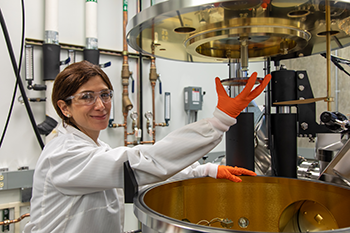
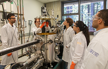
To create the next-generation technology for x-ray imaging, Abbaszadeh and her students will utilize the equipment in her lab at the Baskin School of Engineering to engineer the optical and electrical properties of chalcogenide material – materials that contain one or more chalcogen elements. This facility, which is dedicated to developing detectors based on chalcogenide alloys of the element selenium, is the only such facility in a research setting in the country to Abbaszadeh and Wang’s knowledge.
A dedicated facility for detector development presents a wide range of research opportunities for Abbaszadeh’s lab, as the material has properties that are well-suited for both photodetectors, used for applications ranging from medical imaging to high-energy physics.
This collaborative project will provide exciting opportunities for students in the two researcher’s groups to be part of a larger learning environment in which they can visit each other's labs and gain experience with all facets of their technology, including hardware engineering, AI development, and the clinical setting in which their work will be put to use.
Wang and Abbaszadeh will also collaborate with researchers at the Stanford School of Medicine, who will provide input on how to best design their systems for use in the clinical setting and provide images from real instances of CAC to train Wang’s AI models. Former Stanford instructor Martin Willemink and Stanford Professor Dominik Fleischmann provided input into the development of the project, and Fleischmann, who is the director of Stanford’s cardiovascular imaging division, will continue to lend his expertise. They will also collaborate with industry partners to demonstrate the performance of the new system.
“Between the detector physics that Shiva’s working on, and the artificial intelligence algorithms we're developing at Stanford, we’re providing better image input information but also we will have algorithms that automatically detect and quantify how much calcium there is,” Wang said. “It’s an improvement on multiple fronts.”