Campus News
Tracing neurons in the visual cortex sheds light on brain’s circuitry
Researchers used a combination of techniques to identify different neuronal cell types and trace their connectivity within the mouse visual cortex.
In their efforts to understand how the brain processes information, neuroscientists are trying to identify all of the different types of neurons in the brain and how they connect with one another. Genomic tools have been used to classify neuronal cell types according to their gene expression profiles, but a new study shows that gene expression alone cannot identify all of the different types of neurons.
“Gene expression analysis is not sufficient to define the cell types. Using anatomical methods, we find cell types that are functionally very different within the same genomic cluster,” said Euiseok Kim, assistant professor of molecular, cell, and developmental biology at UC Santa Cruz and first author of the study, published May 11 in Neuron.
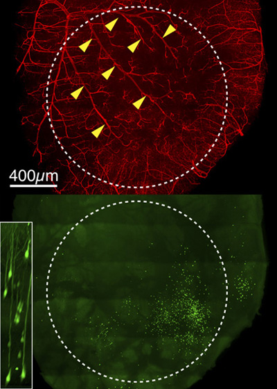
The study focused on neurons that form connections between different visual areas of the cortex in mice. Neurons called cortico-cortical projection neurons relay signals from the primary visual cortex to higher visual areas that play different roles in the processing of visual information. In addition to these “feed-forward” signals, there are also feedback signals from the higher visual areas to the primary visual cortex.
“Within the mouse visual cortices, there are at least ten different areas with different functions, and these areas need to talk to each other to produce the final visual perception,” Kim explained. “We don’t fully understand how they connect to each other, though, because the neurons are so intermingled in the brain that their connectivity is hidden. Only cellular-level dissection can help us see the connections.”
Neuroscientists have developed techniques to trace neural connections using modified viruses, such as an attenuated strain of the rabies virus engineered to infect a specific population of neurons and express a green fluorescent protein. The virus is able to spread across the synapse where one neuron connects to another only once, revealing the direct connections.
“When we examine the brain, we can then see the connected network,” Kim said.
This technique enabled Kim to show that neurons that project from the primary visual cortex to different higher cortical areas differ systematically in their gene expression patterns, even though gene expression analysis alone identifies them as the same genetic type.
Another important finding is that these neurons receive feedback preferentially from the same areas to which they project. This was not unexpected, but it had not been confirmed prior to this study.
“It shows reciprocal connectivity, which is important for understanding the functional role of cortical feedback circuits,” Kim said.
Kim conducted the study with colleagues at the Salk Institute for Biological Studies in La Jolla, California, where he was a postdoctoral researcher prior to joining the faculty at UC Santa Cruz. This work was supported by the National Institutes of Health and the Brain Research Foundation.