Student Experience
Neuroimaging workshop brings leading scientists to UC Santa Cruz
Graduate students and early-career researchers joined scientists from around the world at UC Santa Cruz for a hands-on neuroimaging workshop, gaining practical experience with advanced tools and insight into how researchers study the brain.
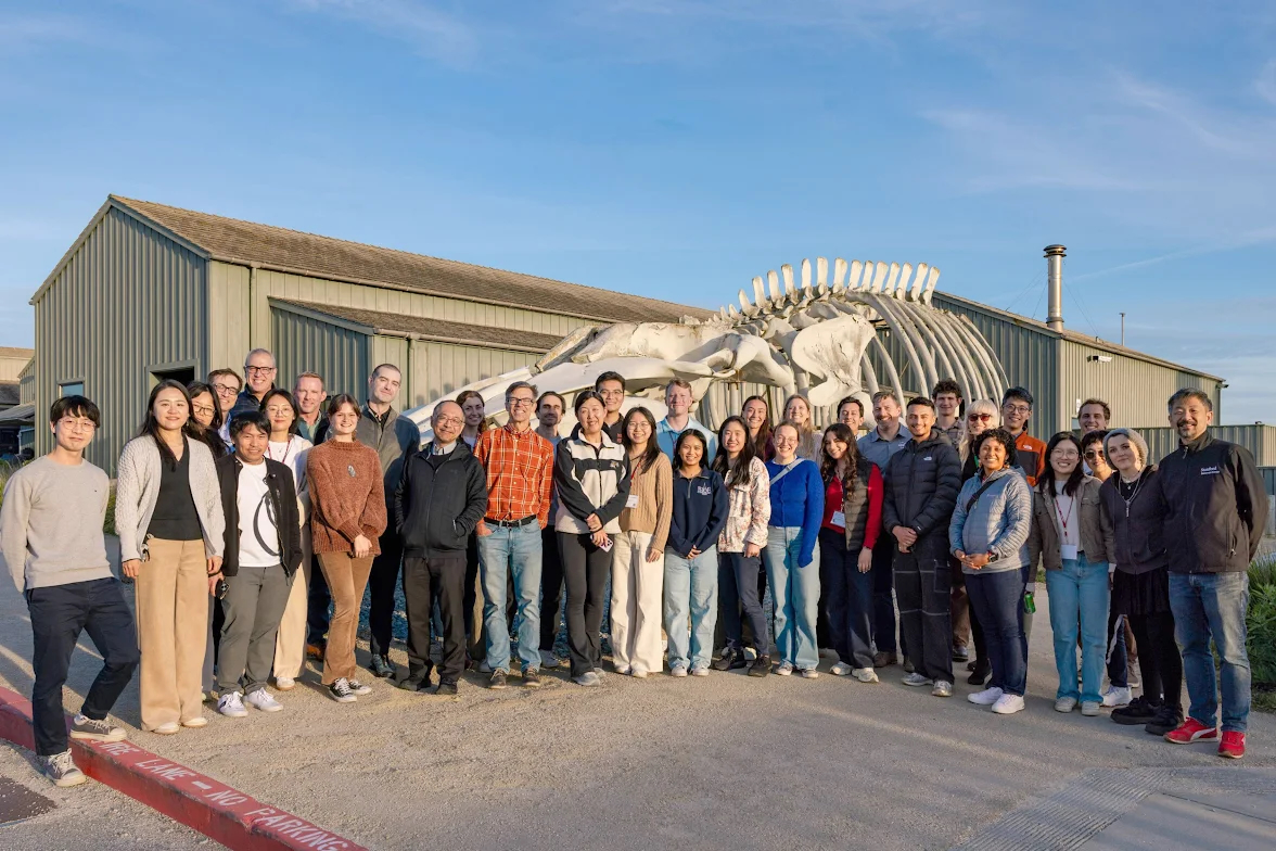
Advanced Techniques in Neuroimaging Workshop participants at the Coastal Science Campus, UC Santa Cruz.
Scientists from around the world came to UC Santa Cruz in April for two days as participants in the Advanced Techniques in Neuroimaging Workshop, a five-day program co-hosted by UC Santa Cruz and Stanford University.
Led by Yi Zuo, professor of molecular, cell, and developmental biology at UC Santa Cruz, and Gordon Wang, associate professor in the Department of Psychiatry at Stanford, the immersive workshop brought together graduate students, postdoctoral researchers, and faculty from Europe, North America, and Asia to learn about cutting-edge neuroimaging tools and techniques.
Throughout the week, participants engaged in demonstrations exploring topics ranging from two-photon microscopy and FRET/FLIM imaging to super-resolution and volumetric imaging techniques that enable the study of neuronal structure and function across scales, as well as a mix of lectures and learning sessions.
For the UC Santa Cruz portion of the workshop, a key highlight was the opportunity to observe live imaging of mouse brains, an area in which Zuo is a global leader. Demonstrations were designed to leverage the infrastructure and unique research capabilities of the Zuo Lab, where advanced imaging technologies were used in real time. UC Santa Cruz was uniquely positioned to host this portion of the workshop since the demonstrations require live animal models and highly specialized lab environments.
“Our goal is to both teach the latest imaging technologies and to create an environment where scientists can see how these tools are actually applied to answer fundamental questions about the brain,” said Zuo. “By combining lectures with demonstrations, we hope to lower the barrier for adopting these approaches and to foster collaborations that extend well beyond the workshop.”
Throughout the two days, internationally recognized leaders in the field held lectures and demonstrations. Among them was Stefan Hell, a Nobel Prize–winning scientist from Max Planck Institutes in Göttingen and Heidelberg, whose groundbreaking work overcame the long-standing diffraction limit in light microscopy. His development of super-resolution fluorescence microscopy transformed the field, enabling researchers to visualize cellular structures at the nanometer scale.
“Our goal is to both teach the latest imaging technologies and to create an environment where scientists can see how these tools are actually applied to answer fundamental questions about the brain.”
Yi Zuo
professor of molecular, cell, and developmental biology at UC Santa Cruz
Zuo also delivered a lecture on confocal and two-photon microscopy, emphasizing in vivo applications of two-photon imaging to study both structural and functional dynamics in the brain, including dendritic spine remodeling and neural activity measurements in behaving animals. Ryohei Yasuda, scientific director at the Max Planck Florida Institute for Neuroscience, also presented. His lab has pioneered techniques to image protein activity within single dendritic spines, offering unprecedented insight into synaptic function at the molecular level.
Additional speakers included Logan Grosenick of Cornell University, who develops computational and imaging approaches to study neural circuits, and Bo Huang of UC San Francisco, whose work focuses on advancing super-resolution microscopy to map subcellular structures and biomolecular organization.
The workshop brought together local, national, and international participants from universities, institutes, and biotechs, including UC Santa Cruz, Stanford, UC San Francisco, Cornell, Max Planx Institutes, and other leading research organizations.