A new cryo-electron microscopy facility is nearing completion at UC Santa Cruz, giving researchers powerful tools for studying the structures and functions of complex molecules involved in human health and disease.
Grants from the National Institutes of Health (NIH) funded the acquisition of a new electron microscope and camera needed to perform cryogenic electron microscopy (cryo-EM), a revolutionary technique for determining the 3-dimensional shapes of large proteins and nucleic acids and the complex assemblies of those molecules that drive the vital functions of living cells.
“A growing community of structural biologists at UCSC is excited to exploit the potential of this technology,” said Melissa Jurica, professor of molecular, cell, and developmental (MCD) biology at UC Santa Cruz.
Structural biologists in several departments at UCSC are doing important work to understand the molecular machinery that operates in the cells of our bodies and underlies human health and disease. This includes research on structures such as the ribosome, spliceosome, cell-cycle regulators, circadian clocks, immune response regulators, childhood viruses, and more.
High-end instruments
Jurica set up UCSC’s first electron microscope with cryogenic capabilities when she joined the faculty in 2003. Cryo-EM technology has improved greatly since then, and in recent years, UCSC researchers have had to go to other institutions to use the latest high-end instruments. Now, they can do the work here and train their students to use state-of-the-art equipment.
“Cryo-EM has gone through a revolution in the past five years or so, made possible by improvements in the technology of the microscopes and the cameras,” Jurica said. “UCSC has a very strong group of structural biologists who need this technology. We all worked together to build this facility.”
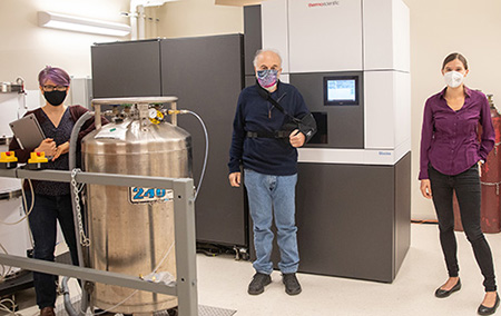
Jurica led the application for the $1.6 million NIH High-End Instrumentation grant that funded the purchase of the new cryo-electron microscope, which includes an “autoloader” device that loads the samples and vastly increases both through-put and stability of the instrument.
Seth Rubin, professor of chemistry and biochemistry, and Harry Noller, professor emeritus of MCD biology, got a separate NIH grant to purchase the specialized camera, a direct electron detector, which is a critical component for achieving high-resolution images of molecular structures. The campus administration, meanwhile, funded renovations for the new facility in the Sinsheimer Laboratories building.
“We’re now in a position to do a lot of really good work,” Jurica said.
Users of the facility will include nearly a dozen faculty members in the Departments of Chemistry and Biochemistry, MCD Biology, Biomolecular Engineering, and Microbiology and Environmental Toxicology. The new facility was crucial to the hiring of a new faculty member, Assistant Professor of Chemistry and Biochemistry Sarah Loerch, who uses cutting-edge cryo-EM methods to study the structure and function of RNA-protein complexes. Focusing on specialized ribosomal complexes, the Loerch lab is working to understand how a cell instructs ribosomes to make the right protein at the right time and in the right place.
Crystallography
Traditionally, the preferred technique for determining the structures of complex biomolecules has been x-ray crystallography. This was the method used by Noller and others to determine the structure of the ribosome, a molecular machine composed of multiple proteins and RNAs that performs protein synthesis in all cells. But x-ray crystallography requires crystallizing the structures first, an extremely challenging step for many biological molecules.
Cryo-EM doesn’t require crystals, and its use has grown dramatically as the methods and equipment have improved. The technique involves flash-freezing samples to capture flexible and dynamic biological molecules in static poses. “With cryo-EM, we can now get direct, high-resolution images of molecules that were not possible to crystallize,” Jurica said.
Noller plans to use cryo-EM images of a ribosome at different steps of protein synthesis to create a molecular movie of the process. With x-ray crystallography, this would require many months of trying to get a crystal for each frame of the movie, but cryo-EM makes it much faster and easier to do. Loerch plans to take the technology one step further and actually image ribosomes while they are inside a neuron to determine how the ribosomes are instructed to make proteins in response to a synapse firing.
Jurica’s lab will be looking at the splicing machinery that edits the RNAs copied from genes; Rebecca Dubois, associate professor of biomolecular engineering, will be looking at the structure of viruses to aid in vaccine development; and Carrie Partch, professor of chemistry and biochemistry, will be using cryo-EM to look at how all the parts of the circadian clock work together to maintain the daily rhythms of our cells.
All of these molecules are large and change their shapes constantly as they do their cellular jobs, which means crystallography would take decades, and might never work. “Cryo-EM lets us take a short-cut straight to the molecules,” Jurica said.
Support from multiple departments and divisions, as well as from campus administration, enabled UCSC to build the new cryo-EM facility, she said. The new facility will open in the new year.