Campus News
A little squid and its glowing bacteria yield new clues to symbiotic relationships
A small molecule produced by bioluminescent bacteria as they colonize the light organ of the Hawaiian bobtail squid may play a key role in establishing the symbiosis.
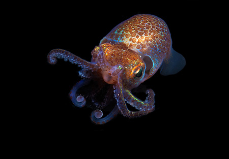
Found hunting at night, the Hawaiian Bobtail squid, Euprymna scolopes displays amazing colors through its light emitting organs. They are common in sand flats around the Hawaiian islands.
The relationship between the Hawaiian bobtail squid and the bioluminescent bacteria living in its light organ has been studied for decades as a model of symbiosis. Now researchers have used a powerful chemical analysis tool to identify a small molecule produced by the bacteria that appears to play an important role in their colonization of the light organ.
The study, published March 9 in the journal mBio, adds a new wrinkle to scientists’ understanding of the chemical signaling involved in this iconic symbiotic relationship. “It’s exciting that there are still new things to discover, even in such a well-studied system,” said corresponding author Laura Sanchez, associate professor of chemistry and biochemistry at UC Santa Cruz.
The Hawaiian bobtail squid is a small nocturnal squid, about the size of a thumb, that lives in shallow coastal waters, hiding in the sand during the day and coming out at night to hunt for small shrimp and other prey. The bioluminescent glow from its light organ is directed downward and adjusted to match the intensity of light from the moon and stars, eliminating the squid’s shadow and masking its silhouette. This “counterillumination” strategy helps conceal the squid both from bottom-dwelling predators and from its own prey.
A juvenile bobtail squid is completely free of bacteria when it first hatches, but within hours its light organ becomes colonized by a very specific type of bacteria called Vibrio fischeri. The baby squid enters an environment teeming with thousands of kinds of bacteria and millions of bacterial cells per milliliter of seawater, of which only a tiny fraction are V. fischeri. Yet only those specially adapted bacteria are able to take up residence inside the light organ.
“It’s a very elegant symbiosis,” Sanchez said. “We already knew that not all strains of V. fischeri are the same—some are better colonizers than others—and we wanted to know if that’s being determined by chemical signals.”
Sanchez’s lab uses a technique called imaging mass spectrometry, which allows researchers to directly visualize the spatial distribution of all kinds of molecules in a sample, such as a squid specimen or a bacterial colony. Most techniques for seeing where specific molecules are in a sample involve labeling the targeted molecules. But imaging mass spectrometry allows untargeted investigations—in other words, you don’t have to know what you’re looking for.
“It’s very difficult to visualize the chemistry in an organism,” Sanchez explained. “With imaging mass spectrometry, we are directly detecting the chemicals, and we know where in the sample they are.”
The small molecule identified in this study is a type of diketopiperazine (DKP), a large family of cyclic dipeptides. This particular DKP—cyclo(d-histidyl-l-proline), or cHP-3—was directly detected in the light organs of the colonized squid. It was also produced more abundantly by strains of V. fischeri that showed increased biofilm formation, which correlates with colonization ability. And finally, supplementing bacterial cultures with cHP-3 led to a concentration-dependent increase in bioluminescence.
“We know that it is produced during the first few hours of colonization when the symbiosis gets established, and we also know that it influences bacterial luminescence, and bioluminescence and colonization are tied together,” Sanchez said.
The results indicate that cHP-3 is an important chemical signal specific to this symbiosis, but the researchers have not yet determined exactly what its role is or the details of its interactions.
“We’re working on that now. We don’t know the mechanisms involved, but there’s a lot more going on than we thought there was,” Sanchez said. “The next steps for us are to find the gene cluster that produces it, and to find how widely used it is.”
In addition to Sanchez, the coauthors of the paper include researchers at the University of Illinois at Chicago and the University of Wisconsin, Madison. This work was supported by the National Institutes of Health and the Chicago Biomedical Consortium.